Multicolor two-photon light-sheet microscopy: fast and colorful
A multidisciplinary group of researchers at the Laboratory for Optics and Biosciences (École Polytechnique-CNRS-Inserm, Palaiseau, France), and at the Institute of Genetics and Molecular and Cellular Biology (Univ Strasbourg-CNRS-Inserm, Strasbourg, France) developed a new fluorescence imaging strategy for the fast observation of intact biological tissue with subcellular resolution by combining light-sheet microscopy and multicolor two-photon excitation using mixed wavelengths. This approach enabled them to image the beating heart of a zebrafish embryo and reconstruct three-dimensional cell movements with sufficient spatial and temporal resolution to track individual cells during a cardiac cycle. It now can be applied to the study of heart formation in vertebrates and diseases leading to heart defects. More generally, the work published in Nature Methods journal opens novel perspectives for fast tissue imaging.
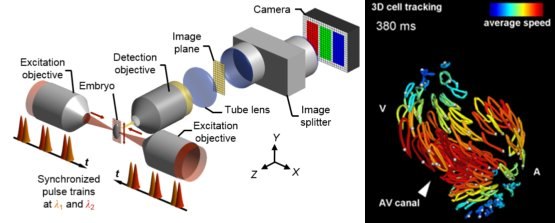
Three-dimensional reconstruction of the beating heart of an embryonic zebrafish using multicolor two-photon light-sheet microscopy. After simultaneously capturing images of blood cells, myocardial cells and cells of the pericardium during a cardiac cycle lasting less than 500 ms, it is possible to follow the trajectories of the individual cells and to quantify their average speed in the atrium (A), the ventricle (V) or the atrio-ventricular canal (AV). Here, a volume of 210 × 230 × 300 μm3 was imaged with 20 ms of acquisition time per image.
© LOB, École polytechnique, CNRS, Inserm / Nature Publishing Group
Publication :
Multicolor two-photon light-sheet microscopy
Pierre Mahou, Julien Vermot, Emmanuel Beaurepaire & Willy Supatto.
Nat. Methods, 11(6), 600-601 (2014).
http://dx.doi.org/10.1038/nmeth.2963