Technologies, setups and developments


Project timeline
Equipment phase: 2013-2023
Running phase: >2015
Microscopy methods development
Tissue imaging:
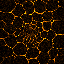
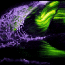
Multimodal multiphoton microscopy: multicolor 2PEF, SHG, THG, 3PEF, CARS, FLIM, polarimetry.
Adaptive optics, photomanipulation.
Fast imaging:
One-photon and two-photon light-sheet (SPIM) microscopy
Super-resolution imaging:
Structured illumination (SIM) / Total internal reflection fluorescence (TIRF)
Soft X-ray / XUV imaging
Some pilot applications (see also Publications)
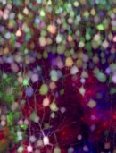
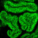
Analyzing lineage & connectivity in the developing brain using brainbow strategies (X-LOB / Inst Vision)
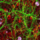
SHG-based analysis of extracellular matrix remodeling and tissue mechanical properties (X-LOB / X-LMS, UPMC)
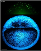
Left-right asymmetry in zebrafish embryos (X-LOB / IGBMC Strasbourg)
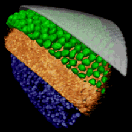
A comparative approach of tissue biomechanics in chordates gastrulation (BioEmergences)
Comparing lineage trees and cell morphodynamics in normal and cloned rabbit embryos (BioEm / INRA Jouy)
Role of the Arp2/3 complex and its regulators in cell morphogenesis, migration and chemotaxis (X-LOB / X-BIOC)
...
Methods developing partners
Polytechnique - Lab for Optics and Biosciences, Palaiseau (coordinator).
Institut d’Optique Graduate School - LCFIO, Palaiseau.
Synchrotron Soleil, Gif/Yvette.
ENS Chimie, Paris.
CNRS CRBM, Montpellier.
I2BC / Imagerie-Gif Light microscopy facility, Gif/Yvette.