Cilia imaging and left-right symmetry breaking
Our web site has moved to this address: https://lob.ip-paris.fr
We investigate left-right symmetry breaking in early zebrafish embryos by devising new in vivo imaging strategies to quantify cilia dynamics and cilia-driven fluid flows. By combining advanced optical tools such as multiphoton microscopy or femtosecond laser ablation with image analysis, we quantify cilia and fluid flow biophysical features in the zebrafish left-right organizer (a.k.a. the Kupffer's vesicle). We use such in vivo experimental data to seed simulations and models and address flow generation and sensing during left-right axis specification.

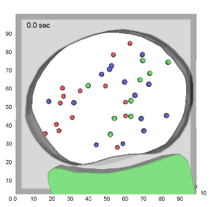
Quantification of the flow surrounding a single beating cilium (up) and within the entire Kupffer's vesicle (down). Adapted from Supatto & Vermot 2011 and Supatto et al, 2008.
Related publications
Chiral cilia orientation in the left-right organizer.
Ferreira RR, Pakula G, Klaeyle L, Fukui H, Vilfan A, Supatto W, and Vermot J
Cell Reports (2018)
Physical limits of flow sensing in the left-right organizer.
Ferreira RR, Vilfan A, Jülicher F, Supatto W*, and Vermot J* (*equal contributors)
eLife (2017)
From cilia hydrodynamics to zebrafish embryonic development.
Supatto W, and Vermot J
Current Topics in Developmental Biology (2011)
An all-optical approach for probing microscopic flows in living embryos.
Supatto W, Fraser SE, and Vermot J
Biophysical Journal (2008)
Research highlights
"Going with the flow": News in Analytical Chemistry (2008)
"4D-particle tracking": Bitplane Learning Center